Diagnostic Over Therapeutic
 Brown SPH has a long and distinguished history as a collaborator in this field. Constantine Gatsonis, professor of biostatistics, joined the American College of Radiology Imaging Network (ACRIN) upon its formation in 1999 as a statistician. Funded by the National Cancer Institute, with an initial grant of $23 million over five years, ACRIN was one of the first national, multi-center research groups focused on diagnostic, rather than therapeutic, medical technologies.
In addition to advances in imaging technology, ACRIN took advantage of other key technological developments, becoming one of the first multi-center studies to collect almost all its data through the new technology of the internet. ACRIN’s interdisciplinary leadership included medical, surgical, and radiation oncologists, patient advocates, and a statistician. Rather than targeting a specific type of cancer, ACRIN investigated the potential of imaging analysis to screen, detect, diagnose and treat cancers across the board.
ACRIN merged with Eastern Cooperative Oncology Group to form ECOG-ACRIN in 2012, the same year that researchers coined the term “radiomics” to describe the extraction of biologically relevant quantitative features from radiological images. It now comprises 1300 member institutions and around 15,000 physicians and research professionals.
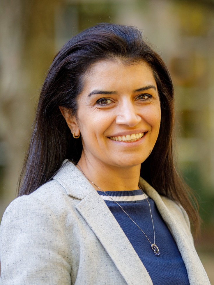
Fenghai Duan, associate professor of biostatistics, who joined the Department’s research track in 2008, does most of his research within ECOG-ACRIN. Lung cancer kills more people worldwide than any other cancer. As part of the National Lung Screening Trial (NLST) research team, Duan’s work analyzing the success of screening for lung cancer—which using either X-rays or CT (computed tomography) demonstrated that the sensitivity of CT scans allowed for earlier detection and earlier treatment, leading to a twenty percent relative reduction in lung cancer specific mortality compared to X-rays, leading in 2011 to an immediate change in national screening guidelines.
But both CT scans and X-rays produce a very high number of “false positives,” turning up nodules that more than 90% of the time turn out to be benign and pose little health threat, which is why screening guidelines depend on risk factors—mainly age and smoking history. Public health policymakers must balance the harm of false positives—including anxiety and unnecessary surgery—with the harm of failing to detect cancer until it is too late to treat. Duan and other researchers began to not just look for nodules, but to analyze the features of the nodules they found—size, location, shape, and so on. By connecting these data to patient clinical history and outcomes, Duan and his collaborators developed a pulmonary nodule risk prediction model that could distinguish with much greater accuracy which nodules would develop into full-blown cancer.
But the predictive power of this model was limited by the data on which it was based, and the computing power to process those data. As computer power has increased, so has the quality of radiologic imaging, with increasing resolution producing images of greater and greater granularity. “Rather than just a few features of the nodule, radiologists can now provide hundreds or even thousands of imaging features,” Duan said. “That’s what we call radiomics: massive quantitative features associated with an image of the nodule.” Radiomics harnesses this fount of new data with the new techniques of machine learning, yielding vastly more complex and accurate prognostic tools. In the last two years, Duan has shown that the radiomics-based model he has developed in collaboration with pulmonologists and radiologists is better at distinguishing benign from malignant nodules than the standard of care risk assessment.


 Brown SPH has a long and distinguished history as a collaborator in this field. Constantine Gatsonis, professor of biostatistics, joined the American College of Radiology Imaging Network (ACRIN) upon its formation in 1999 as a statistician. Funded by the National Cancer Institute, with an initial grant of $23 million over five years, ACRIN was one of the first national, multi-center research groups focused on diagnostic, rather than therapeutic, medical technologies.

